Anal papillitis
Anal papilla – papillitis
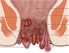
At the bottom of the ampoule wall of the rectum are called morganievy columns and crypts. These formations, as it were surrounded by semilunar valves. Sometimes the free edge of the valve may be a small triangular elevation – is anal papillae. These papillae, if their size is less than 1 cm in diameter and they are at the level of the anorectal line – this is the norm.
To identify these papillae doctor can by digital rectal examination. However, they are defined as painless nodules in the upper third of the anal canal. During the anal papillae anoscopies have a pale pink color.
The form of the anal papillae can be of two kinds:
The first kind – nipples have a wide base and a triangular shape,
The second kind – nipples have a narrow foot, and a spherical shape.
In the case of inflammation of the anal papillae, which usually occurs when they are hypertrophied, developed papillitis. Usually, the infection spreads from the papillomas in the inflamed morganievyh crypts, or when proctitis.
In papillitis is usually marked swelling of the tissues of the papilla, its redness (hyperemia), and soreness. If there is a long-term trauma of inflamed papillae, for example, hardened feces, or foreign bodies, its tip may be ulcerated.
Reasons papillitis
Papillitis – an inflammation of the anal papillae. Inflammation can usually occur in the hypertrophied papillae, where they can fall out of their rectum out. Sometimes the size of fallen buds can reach 3 – 4 cm in diameter.
Among the causes of hypertrophy of anal papillae may be noted:
- Mechanical injury and chemical irritation of the papillae stool – how dense and, conversely, liquid.
- Poor circulation in the pelvis, namely – poor blood flow that occurs with hemorrhoids.
- Inflammatory diseases of the anal canal. Main role is given kriptitu. Therefore, the presence of hypertrophied anal papillae are often evidence of inflammation in the anal canal.
Diagnosis papillitis
First of all, the doctor asks the patient and collects his complaints, as well as history (how the disease began, with which it began to appear, etc.)
An important role in the diagnosis of papillita plays examination of the anus, both at rest and during straining the patient (to see whether the buds do not fall). In addition, using digital rectal examination, and, anoscopy, and sigmoidoscopy. These methods allow the doctor to see the status of the anal canal and rectum, reveal the presence of anal papillae, kriptita and other related diseases.
Differential diagnosis papillitis
Papillita Symptoms can occur in other diseases of the rectum, and the hypertrophied papillae themselves to be, above all, be distinguished from polyps of the colon.
 In some cases, doctors can take a normal anal papillae during their hypertrophy of polyp of the rectum or anal canal polyp. Indeed, during hypertrophy anal papillae become “foot”, and then they are his views are similar to polyp. However, the true polyps are always located above the anorectal line. Polyp of the rectum is called adenomatous structure and is usually covered by a single layer of columnar epithelium. By the color of the polyp of the rectum is the same as the normal intestinal mucosa.
Unlike polyps, anal papillae are located in the anal canal at the anorectal line and consist of collagen fibers with a small amount of adipose tissue. These anal papillae are covered with stratified squamous epithelium, and their color – white.
In some cases, hypertrophied anal papillae must be distinguished from the guard bumps that occur in patients with chronic anal fissure, as well as chronic hemorrhoids. Typically, the guard bumps are located at the upper or lower edge of anal fissure.
In contrast to the hypertrophied anal papillae, internal hemorrhoids are usually above the anorectal line, the color of dark red, according to their consistency they are soft to the touch (no thrombosis).reatment papillitis
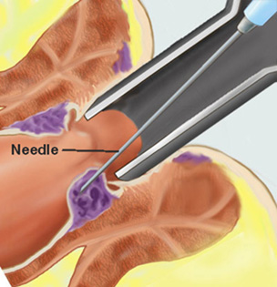
If you find a doctor anal papillae, it is important to understand that they are the norm. If the buds do not cause any anxiety the patient (rectal prolapse, pain) then they should not be touched. If they cause discomfort to the patient, then they are usually excised, usually with the crypt and the semilunar valve. Surgery to remove the anal papilla is usually performed under local anesthesia.
In the case of papillita conservative therapy is usually performed as in the crypt, and the removal of the papilla is performed only after the acute inflammation subsided.
By themselves, hypertrophy and inflammation of the anal papillae are not a serious pathology, but it is important to know that inflammation of the structures of the rectum is never isolated and always associated with something. Therefore, you should look for the cause of inflammation of the anal papilla.
Our Expertise in Medical Services
Rana Hospital was started with a dream of providing the best facilities for Piles, Fissures, Fistula and all Anorectal problems.

Mon to Sat:
8:30 AM – 1:30 PM
4:00 PM – 6:30 PM
Mon to Sat:
9:00 AM – 1:30 PM
4:00 PM – 6:30 PM
Make an Appointment
 Accreditation & Awards
The Hospital organized a free checkup camp for PILES on 24th October 2013 in which 717 patients from all over India were examined. It was followed by one day free operation camp on 27th October 2013. Dr Suri single-handedly performed 391 free surgeries in 8 hours & 45 minutes setting a World Record.


